


卵母细胞的生发泡破裂(Germinal vesicle breakdown,GVBD)通常被认为是卵母细胞成熟的重要标志[16]。大部分海水贝类解剖卵母细胞的生发泡(GV)在自然海水中不会发生破裂,只有采用维生素、氨海水、5-羟色胺(5-HT)、多巴胺、组胺等化学药物浸泡卵母细胞时,生发泡才会破裂[17⇓⇓-20]。5-HT广泛存在于海水贝类的神经系统和性腺组织中[21],在海水贝类繁殖过程中起着非常重要的作用,特别是在促进卵母细胞成熟等方面[22]。Guerrier P等[23]研究表明,5-HT可以促使菲律宾蛤仔(Ruditapes phillippinarum)解剖卵母细胞的生发泡破裂;Gloria M等[24]研究表明,采用5-HT浸泡可以促熟紫扇贝(Argopecten purpuratus)的解剖卵母细胞成熟,为自交和杂交试验的开展提供良好基础。氨海水在刺激海水贝类排精产卵、促进精卵成熟等方面起重要作用[25]。喻达辉等[26]研究了氨海水对合浦珠母贝(Pinctada martensii)解剖精卵的体外促熟作用,结果表明氨海水对合浦珠母贝的精子起激活作用,主要成分是NH4+;李浩浩等[27]对栉江珧(Atrina pectinata)解剖卵母细胞体外促熟的研究结果表明,采用一定浓度的氨海水浸泡可以明显提高卵母细胞的GVBD发生率和受精率,且受精卵在发育过程中畸形率较低。本研究以钝缀锦蛤为研究对象,采用了不同浓度的5-HT、氨海水在不同时间下浸泡钝缀锦蛤卵母细胞,通过观察GVBD、受精率和胚胎发育情况,从而获得5-HT、氨海水对钝缀锦蛤解剖卵母细胞的最佳体外促熟参数,为海水贝类卵母细胞体外促熟及遗传选育、多倍体育种提供基础资料。
1 材料与方法
1.1 精、卵的获得
实验用钝缀锦蛤为厦门市翔安区大嶝街道小嶝池塘养殖的2龄亲贝,平均体质量为(35.77±7.40)g,平均壳长为(62.44±3.91)mm,平均壳高为(40.53±2.34)mm,平均壳宽为(26.08±1.65)mm。钝缀锦蛤亲贝经过池塘暂养1个月后,从中挑选性腺成熟度好的个体,解剖获得精、卵。卵液用300目筛绢网过滤去除较大组织等,再用500目筛绢网收集卵子等。精液用500目筛绢网过滤杂质。检查卵子成熟度及精子活力等情况,并选择成熟度高的卵子及活力好的精子用于开展实验。
1.2 解剖卵母细胞的体外促熟
在室温(22℃)条件下,在50 mL离心管中分别加入30 mL成熟度高的卵液,然后加入不同浓度的5-HT和氨海水处理液,卵母细胞密度保持在500~1 000个/mL,混匀后开始计时。5-HT处理浓度梯度设定为0.01、0.1、1、10、100、1 000 μmol/L;氨海水处理浓度梯度设定为0.002 5%、 0.005%、 0.010%、 0.015%、 0.020%和0.025%。药品处理时间梯度分别为15、30、45、60 min。采用在自然海水中浸泡的卵母细胞作为对照组,每次实验设置3个平行组。
1.3 人工授精
用500目筛绢网过滤去除处理液,将卵子收集在500 mL烧杯中,然后加入2~3滴活力好的精子,充分混匀。在受精过程中,应尽量搅动受精卵液,保证溶解氧充足。
1.4 观察记录GVBD发生率和受精率
受精卵发育2 h后,取样进行显微镜拍照计数,同时计算GVBD发生率和受精率。
1.5 数据处理
实验数据使用SPSS 19.0软件进行单因素方差分析,采用Duncan’s多重比较检验不同组差异显著性,显著水平设置为P<0.05。
2 结果与分析
2.1 钝缀锦蛤卵母细胞体外促熟的形态变化
图1
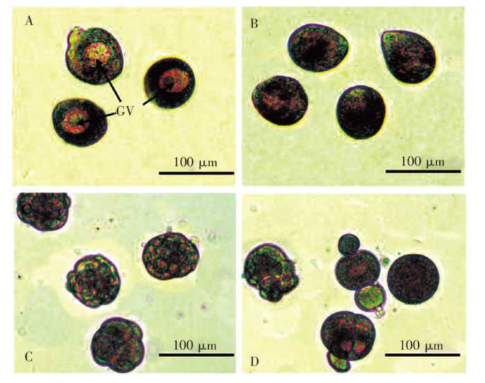
图1
钝缀锦蛤解剖卵子的形态变化
注:A.生发泡未破裂的卵细胞;B.生发泡破裂的卵细胞;C.正常发育的受精卵;D.畸形受精卵。
Fig.1
Morphology changes of oocyte stripped from T.conspersus
Notes:A.Oocytes with germinal vesicle;B.GVBD of oocytes;C.The normal fertilized egg;D.Malformation.
2.2 5-HT对钝缀锦蛤卵母细胞的促熟作用
钝缀锦蛤解剖卵母细胞在正常海水中浸泡一段时间后,生发泡几乎不会发生破裂;采用5-HT浸泡可以显著促使钝缀锦蛤卵母细胞的成熟(图2)。当处理时间相同时,采用10 μmol/L~1 mmol/L 5-HT 浸泡钝缀锦蛤卵母细胞,GVBD发生率差异不显著(P>0.05);采用0.01 ~1 μmol/L 5-HT 浸泡钝缀锦蛤卵母细胞,GVBD发生率差异显著(P <0.05),表现出一定的浓度依赖关系。除45 min和60 min同一浓度实验组之间差异不显著(P>0.05)外,其他不同时间同一浓度实验组差异显著(P <0.05),表现出一定的时间依赖关系。
图2
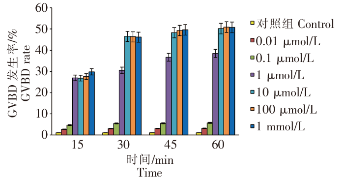
图2
不同浓度 5-HT 对 GVBD 发生率的影响
Fig.2
Effect of different concentration of 5-HT on GVBD rate
钝缀锦蛤的卵母细胞经过不同浓度5-HT浸泡处理后,人工授精的受精率见图3。卵母细胞经过5-HT浸泡后,可以显著提高受精率(P<0.05)。除了0.01~0.1 μmol/L 5-HT实验组外,其他实验组(1 μmol/L~1 mmol/L 5-HT)表现出一定的时间依赖关系,受精率随时间增加而提高。当处理时间相同时,表现出一定的浓度依赖关系,1~10 μmol/L 5-HT实验组的受精率显著高于其他实验组(P<0.05);其中,10 μmol/L 5-HT浸泡60 min实验组受精率最高,达到48.57%。
图3

图3
不同浓度 5-HT 对受精率的影响
Fig.3
Effect of different concentration of 5-HT on fertilizing rate
2.3 氨海水对钝缀锦蛤卵母细胞的促熟作用
对钝缀锦蛤解剖卵母细胞采用一定浓度的氨海水浸泡,可以显著促使卵母细胞的成熟(图4)。当处理时间相同时,采用0.002 5%~0.010%氨海水浸泡钝缀锦蛤卵母细胞,GVBD发生率差异不显著(P>0.05);采用0.015%~0.025%氨海水浸泡钝缀锦蛤卵母细胞,GVBD发生率差异显著(P <0.05),表现出一定的浓度依赖关系。除了0.002 5%氨海水实验组外,其他实验组随着处理时间的增加,GVBD发生率差异显著(P<0.05),表现出一定的时间依赖关系。
图4
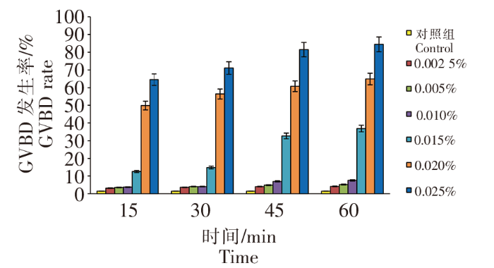
图4
不同浓度氨海水处理对 GVBD 发生率的影响
Fig.4
Effect of different concentration of AS on GVBD rate
钝缀锦蛤的卵母细胞经过不同浓度氨海水浸泡处理后,人工授精的受精率见图5。卵母细胞经过氨海水浸泡后,可以显著提高受精率(P<0.05)。除了0.002 5%~0.005%、0.025%氨海水实验组外,其他实验组(0.015%~ 0.020%氨海水)表现出一定的时间依赖关系,15 min至45 min时间内受精率随时间增加而提高。当处理时间相同时,表现出一定的浓度依赖关系,0.015%~0.025%氨海水实验组的受精率显著高于其他实验组(P<0.05);其中,0.020%氨海水浸泡45 min实验组受精率最高,达到50.75%。
图5
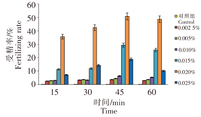
图5
不同浓度氨海水处理对受精率的影响
Fig.5
Effect of different concentration of AS on fertilizing rate
采用不同浓度的氨海水浸泡钝缀锦蛤卵母细胞,虽然可以获得较高GVBD发生率和受精率,但受精卵的畸形率较高,部分受精卵在卵裂过程中就停止胚胎发育。
3 讨论
3.1 5-HT和氨海水对海水贝类卵母细胞促熟作用
在海水贝类人工育苗过程中,亲贝性腺成熟程度非常关键,卵子质量的好坏直接决定整个育苗过程的成功率[28]。海水贝类卵母细胞的形态、GVBD发生率以及细胞器的状态等都是判断卵母细胞成熟度的重要依据[29],因此,在开展钝缀锦蛤解剖卵母细胞的体外促熟过程中,应选择卵径在70~80 μm、形状圆形的卵母细胞。海水贝类的解剖配子存在两种状态:一种是卵生型牡蛎等[30]的解剖卵母细胞可以直接人工授精发育,而绝大部分海水贝类,如菲律宾蛤仔、硬壳蛤(Mercenaria mercenaria)、马氏珠母贝(Pinctada fucata)、栉孔扇贝(Chlamys farrer)等解剖卵母细胞不能直接受精或受精率极低[31],这是因为解剖卵母细胞含有的生发泡在生理上是不成熟的,必须经过一定浓度的药物(5-HT、氨海水、维生素、多巴胺等)浸泡处理后,卵母细胞才能够受精。
本研究采用了5-HT和氨海水浸泡钝缀锦蛤解剖卵母细胞,结果表明5-HT和氨海水均能够促使卵母细胞的生发泡破裂,GVBD发生率达到80%以上,受精率达到45%左右。采用5-HT和氨海水浸泡卵母细胞,钝缀锦蛤与其他海水贝类获得的最高GVBD发生率和受精率各不相同(表1),这可能与不同种类生活习性及生活环境条件(温度、pH等)有关。
表1 海水贝类卵母细胞经5-HT和氨海水浸泡获得的最高GVBD发生率和受精率
Tab.1
| 品种 Varieties | 氨海水 Ammonia seawater | 5-羟色胺 5-hydroxytryptamine(5-HT) | 文献 References | ||
|---|---|---|---|---|---|
| GVBD发生率/% GVBD rate | 受精率/% Fertilization rate | GVBD发生率/% GVBD tate | 受精率/% Fertilization rate | ||
| 栉孔扇贝 Chlamys farrer | 100 | 15.88 | 40 | 24.74 | 邸炜鹏等[19] |
| 马氏珠母贝 Pinctada fucata | - | - | 59 | - | 王琦等[22] |
| 栉江珧 Atrina pectinata | 90 | 88 | 33 | 5 | 李浩浩等[27] |
| 硬壳蛤 Mercenaria mercenaria | - | - | 71.11 | - | 王清等[32] |
| 钝缀锦蛤 Tapes conspersus | 84.44 | 50.75 | 50.86 | 48.57 | 本研究 |
3.2 处理时间和药物浓度对钝缀锦蛤卵母细胞受精发育的影响
邸炜鹏等[19]采用0.005%氨海水处理15 min后、再用2 mmol/L 5-HT处理栉孔扇贝解剖卵子,卵子的受精率达到40.02%。李浩浩等[27]采用一定浓度的氨海水(0.008%~0.014%)浸泡栉江珧解剖卵母细胞40~60 min,可以显著提高GVBD发生率和受精率,受精卵在发育过程中的畸形率较低;而采用5-HT、多巴胺和维生素浸泡卵母细胞,体外促熟效果不明显。本研究表明,1~10 μmol/L 5-HT和0.015%~0.025%氨海水均能够显著提高钝缀锦蛤解剖卵母细胞的GVBD发生率和受精率,0.015%~ 0.025%氨海水实验组的GVBD发生率高于1~10 μmol/L 5-HT实验组,虽然受精率相似,但氨海水实验组的受精卵发育过程中畸形较多;当药物浸泡时间为45~60 min时,GVBD发生率和受精率达到了最大值。因此,对钝缀锦蛤解剖卵母细胞进行体外促熟时,药物浓度及浸泡时间应控制在适宜范围内,从而确保较高的GVBD发生率和受精率。
3.3 解剖授精在海水贝类遗传育种中的作用
目前在海水贝类人工育苗生产中,只有牡蛎等极少数海水贝类采取人工授精方法进行育苗生产,其他海水贝类主要采用人工催产方法进行育苗生产[31]。由于海水贝类是分批成熟、分批产卵的,在人工催产时的产卵不同步、产卵时间长等因素都直接影响育种工作的顺利开展。因此,开展海水贝类卵母细胞的体外促熟方法研究可以解决卵子受精同步性等问题,为海水贝类多倍体育种、选择育种、杂交育种的开展提供有力的技术支撑。
参考文献
盐度、pH、氨氮对钝缀锦蛤稚贝生长及存活的影响
[J].
为探究钝缀锦蛤(Tapes conspersus)稚贝对盐度、pH、氨氮的适应性,本研究在室内可控条件下,分析了不同盐度(10、15、20、25、30、35、40)、pH(2.5、3.0、3.5、4.0、4.5、5.0、5.5、6.0、6.5、7.0、7.5、8.0、8.5、9.0、9.5、10.0)、氨氮(0.5、1、5、10、20、50、100、250 mg/L)对钝缀锦蛤稚贝的生长和存活的影响。不同盐度条件下实验结果表明,钝缀锦蛤稚贝的最适生长和生存盐度为25~35。不同pH条件下实验结果表明,钝缀锦蛤稚贝生存的适宜pH为4.0~8.5,稚贝对低pH耐受性比高pH更强。不同氨氮条件下实验结果表明,氨氮浓度在0.5~10.0 mg/L范围内,稚贝的存活率均为100%;氨氮浓度大于20.0 mg/L时,随着氨氮浓度的升高,稚贝存活率显著下降;在24 h内,不同氨氮浓度下各组稚贝均存活良好,存活率均大于98.33%;48 h、72 h和96 h时,稚贝的半致死浓度分别为123.68、77.36、49.00 mg/L,安全浓度分别为12.37、7.74、4.90 mg/L。
Hatchery production of diploid and triploid clams,Tapes dorsatus(Lamarck 1818):a potential new species for aquaculture
[J].
Salinity studies on the clams Katelysia rhytiphora(Lamy)and Tapes dorsatus(Lamarck)
[J].
Effect of different growing techniques and substrate types on the growth and survival of the clams Tapes dorsatus(Lamarck)and Katelysia rhytiphora(Lamy)
[J].
Mammalian AT2 receptors expressed in Xenopus laevis oocytes couple to endogenous chloride channels and stimulate germinal vesicle break down
[J].
Kinetics and fates of ammonia,urea,and uric acid during oocyte maturation and ontogeny of the Atlantic halibut(Hippoglossus hippoglossus L.)
[J].
Effect of simultaneous variation in temperature and ammonia concentration on percent fertilization and hatching in Crassostrea ariakensis
[J].The combined effects of temperature and ammonia concentration on the percent fertilization and percent hatching in Crassostrea ariakensis were examined under laboratory conditions using the central composite design and response surface methodology. The results indicated: (1) The linear effects of temperature and ammonia concentration on the percent fertilization were significant (P 0.05). (2) The linear effect of temperature on the percent hatching was highly significant (P 0.05). The quadratic effects of temperature and ammonia concentration on the percent hatching were highly significant (P 0.05). Temperature was more important than ammonia in influencing the fertilization and hatching in C. ariakensis. (3) The model equations of the percent fertilization and hatching towards temperature and ammonia concentration were established, with the coefficients of determination R-2=99.4% and 99.76%, respectively. Through the lack-of-fit test, these models were of great adequacy. The predictive coefficients of determination for the two model equations were as high as 94.6% and 98.03%, respectively, showing that they could be used for practical projection. (4) Via the statistical simultaneous optimization technique, the optimal factor level combination, i.e., 25 C-circle/0.038 mg mL(-1),was derived, at which the greatest percent fertilization 95.25% and hatching 83.26% was achieved, with the desirability being 97.81%. Our results may provide advantageous guidelines for the successful reproduction of C ariakensis. (C) 2014 Elsevier Ltd.
Serotonin-like immunoreactivity in the central nervous system and gonad of the scallop,Patinopecten yessoensis
[J].
Evidence for the involvement of internal calcium stores during serotonin -induced meiosisreinitiation in oocytes of bivalve mollusk Ruditapes phillippinarum
[J].In contrast to the situation found in the bivalves Barnea candida and Spisula solidissima, prophase-arrested oocytes of Ruditapes philippinarum cannot be fertilized when removed from the ovary. They must first undergo germinal vesicle breakdown under the influence of the neurohormone serotonin (5-HT), which drives them to a second block occurring in metaphase of the first maturation division. In the studies described in this paper, we investigate the possibility that calcium is involved as a second messenger in controlling this first step in the reinitiation of meiosis. Our data show that, in addition to 5-HT, ionophore, thapsigargin, and the weak bases ammonia and procaine can also induce prophase-arrested oocytes of Ruditapes to resume meiosis. 5-HT, thapsigargin, and ammonia all trigger a surge of intracellular Ca2+ and are effective even in the absence of external Ca2+. That such Ca2+ transients, which are enhanced in the presence of external Ca2+, actually play a key role in the process of meiosis reinitation is shown by the fact that loading the oocytes with BAPTA/AM or treating them with D-600 blocks maturation. In contrast, excess KCl, which has been shown to trigger meiosis reinitiation of prophase-arrested oocytes of Barnea and Spisula and to activate metaphase I-arrested oocytes of Ruditapes, does not produce any significant intracellular Ca2+ transient nor does it reinitiate meiosis, when added to Ruditapes prophase-arrested oocytes. These data suggest that such voltage-operated Ca2+ channels may only appear during the course of maturation and that both intracellular and extracellular Ca2+ are involved in triggering 5-HT-dependent release from the prophase block in this species.
A method to eliminate selffertilization in a simultaneous hermaphrodite scallop.1.Effects on growth and survival of larvae and juveniles
[J].
Gametogenesis of Atrina maura(Bivalve:Pinnidae)under artificial conditions
[J].
Isolation and functional characterization for oocyte maturation and sperm motility of the oocyte maturation arresting factor from the Japanese scallop,Patinopecten yessoensis
[J].In bivalves, serotonin (5-hydroxytriptamine, 5-HT) acts as a major promotional factor in oocyte maturation, sperm motility, and sequential spawning. The previously reported novel neuronal protein, oocyte maturation arresting factor (OMAF) that was found in the central nervous system and hemolymph of the Japanese scallop, Patinopecten yessoensis, has an inhibitory activity in the 5-HT-induced oocyte maturation via a receptor-mediated mechanism, resulting in an arrest of spawning [30]. In this study, OMAF protein was isolated from the supernatant of hemolymph of the scallop using gel and anion-exchange chromatography, and SDS-PAGE. Three digested partial peptides with 4, 11, and 16 amino acid residues were determined through reversed-phase HPLC and amino acid sequencing. The anti-OMAF antibodies generated against the obtained peptides with 11 and 16 amino acid residues were applied to immunohistochemistry and 5-HT-induced spawning and oocyte maturation assays. Fusiform OMAF neurons were localized in the external area of the anterior lobe of the cerebral ganglion, supporting our presumption that OMAF was secreted from the cerebral and pedal ganglia (CPG). Pretreatment with anti-OMAF antibody on three kinds of bivalve species showed a strong in vivo amplification of 5-HT-induced release of egg and sperm, and an in vitro restoration of 5-HT-induced germinal vesicle breakdown (GVBD) from inhibition by the CPG extract, suggesting the release from suppressive activity of OMAF due to the absorption with antibody. These results confirm that the isolated peptides are from OMAF and OMAF acts as an inhibitor of 5-HT-induced oocyte maturation and sperm motility as previously reported.Copyright © 2012 Elsevier Inc. All rights reserved.
